Prof. Ozgun Gokce PhD
Spatial Dynamics of Neurodegeneration
Medical Faculty, UKB, University of Bonn & DZNE
Long before symptoms emerge, neurodegeneration begins with subtle shifts in the state and behavior of individual brain cells. The Gokce Lab traces these changes across molecular, spatial, and structural scales, revealing which genes each cell activates, where it resides in the tissue, and how its ultrastructure changes.
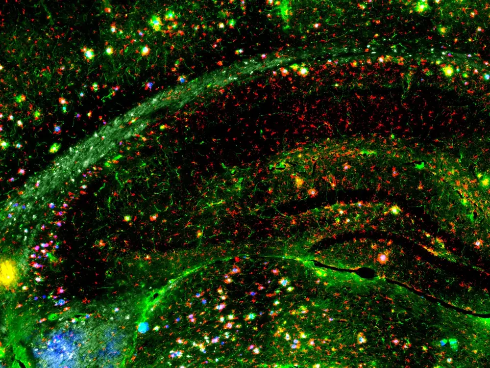
What we do
We map how brain cells change with age and disease. One cell at a time, in place.
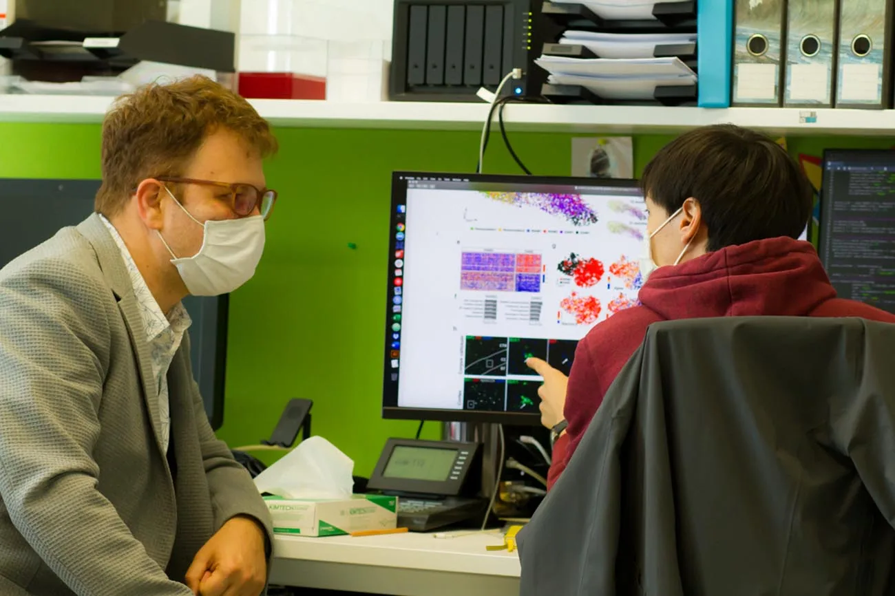
An interdisciplinary group at the University of Bonn and DZNE, where biologists, engineers, physicians, and computational scientists work on the same data.
Single-cell genomics, spatial transcriptomics, electron microscopy, machine learning, and new methods when a question demands them.
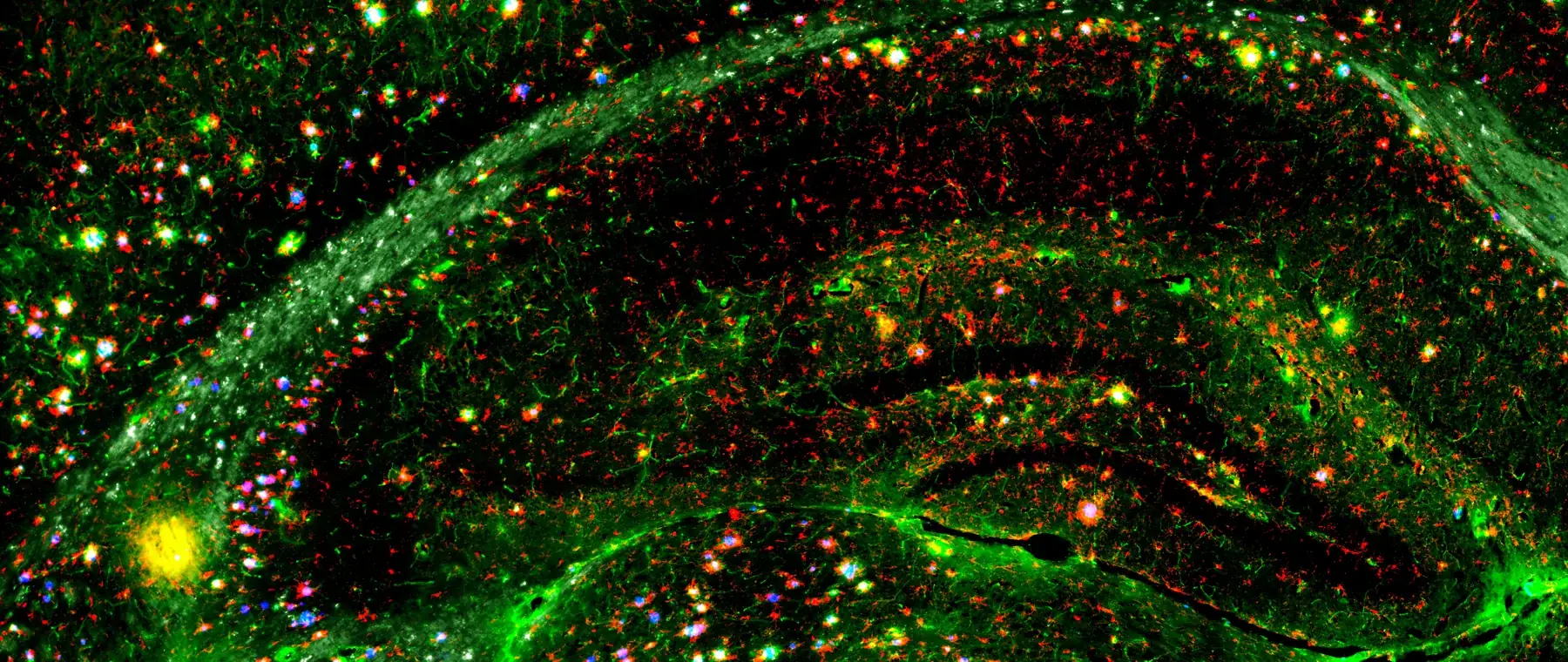
What the data look like
Where this work is published

News
Full news archive-
Three community papers on single-cell and spatial genomics in neuroscience
Ozgun Gokce co-led three companion papers in Nature Neuroscience setting out how single-cell and spatially resolved genomics experiments should be designed, validated and interpreted in the nervous system.
Read the paper -
T cells trigger myelin damage in a mouse model of amyloidosis
Shreeya Kedia, Hao Ji, Peter Androvič, Cem Buğra Kaboğlu and colleagues found that CD8+ T cells drive microglia into a damaging interferon-activated state, and that removing those T cells or blocking the pathway reduces myelin damage in amyloid-depositing mice.
Read the paper -
Skull bone marrow has its own molecular identity
Work with the Ertürk lab found that the marrow inside the skull differs from marrow elsewhere in the body and changes distinctly in neurological disease.
Read the paper
Selected papers
All publications- CD8+ T cells induce interferon-responsive oligodendrocytes and microglia in white matter agingNature Neuroscience · 2022doi.org/10.1038/s41593-022-01183-6
- White matter aging drives microglial diversityNeuron · 2021doi.org/10.1016/j.neuron.2021.01.027
- Spatial transcriptomics-correlated electron microscopy maps transcriptional and ultrastructural responses to brain injuryNature Communications · 2023doi.org/10.1038/s41467-023-39447-9
- T cell-mediated microglial activation triggers myelin pathology in a mouse model of amyloidosisNature Neuroscience · 2024doi.org/10.1038/s41593-024-01682-8
The people behind it
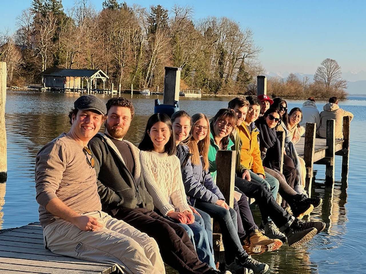
Meet the team
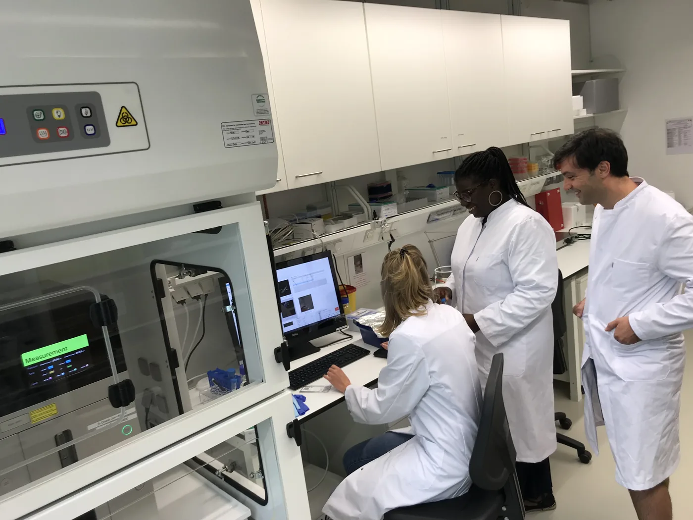
Working here
We take PhD students, postdocs and master's students who want to work at the boundary between experiment and computation. You do not need to arrive fluent in both. You do need to want to be.